- 54 VAS. SOFIAS AVENUE ATHENS 115 28
- (+30) 210 729 75 71
Dysplastic nevi
Dysplastic or atypical nevi, are acquired nevi that present a disorder in their architecture as well as some degree of atypia in their melanocytes. They usually appear in adolescence or later and are quite common in the white race. They have a particular biological behavior and an increased risk for the development of melanoma.
Main risk factors for their development are family history and genetic predisposition, with specific gene mutations being implicated, as well as overexposure to sunlight. The latter seems to be able to affect the number, clinical appearance and progression of atypical nevi, although these nevi can also appear in non-sun-exposed areas of the body.
When more than 20 dysplastic nevi are detected in the same person, we speak of “dysplastic nevus syndrome”. This syndrome can be sporadic or familial and is a risk indicator for the development of melanoma, as it increases the relative risk 4-6 times.
Clinically, dysplastic nevi are usually flat or raised lesions that present irregular borders, color variation, heterogeneity in shape and size >5mm. They can be located anywhere on the body, but mainly on the trunk and extremities.
Based on the well-known ABCDE rule (Asymmetry, Border, Color, Diameter, Evolution), a mole is always evaluated for its symmetry, its outline, the distribution of pigment, its size and its evolution. The earliest sign of transformation of a dysplastic nevus is the modification of its color either by increasing pigmentation or by the development of polychromy. Additional indications for the development of a melanoma are a sudden increase in size, a change in its borders, bleeding or ulceration and symptoms, such as pain or itching.
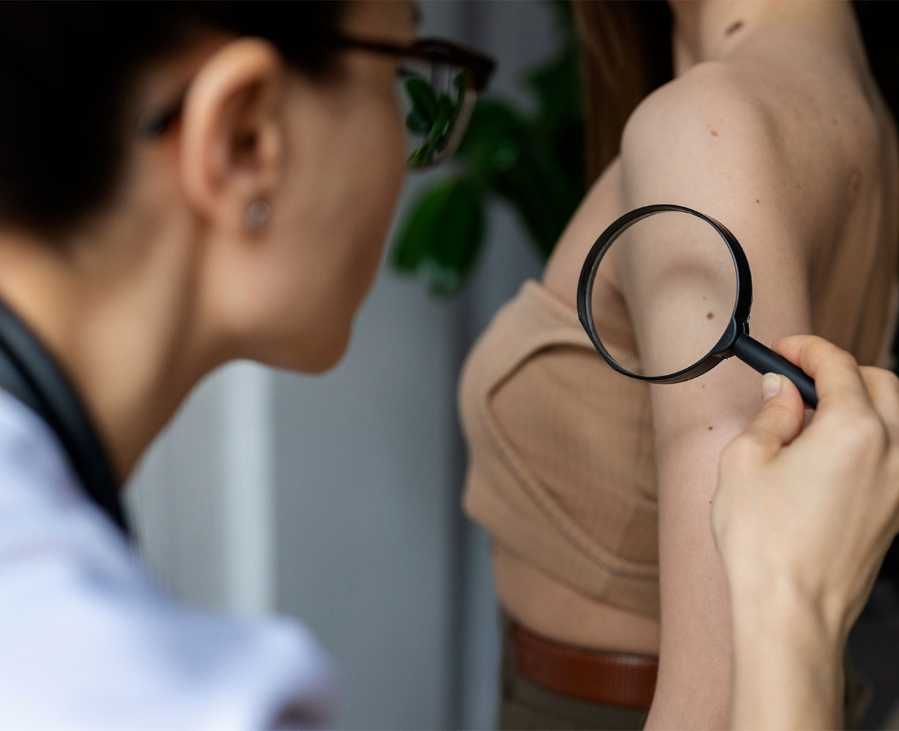
Patients with atypical moles should routinely use a broad-spectrum sunscreen and should avoid, as much as possible, excessive sun exposure and UV-emitting tanning devices (primary prevention). For monitoring, is recommended a complete cutaneous examination with dermoscopy and total body photography at least once a year, as well as educating patients to perform self-examination monthly. The aim of the above is to identify lesions with mild or moderate atypia, so that they can be closely monitored, and to identify lesions with severe atypia early, so that they can be surgically removed and histologically examined (secondary prevention).
The clinical examination is supplemented by taking a detailed personal and family history. Particular emphasis is placed on sun exposure habits, recent changes observed in moles, and the presence of a history of melanoma in both patients and their first- or second-degree relatives.