- 54 VAS. SOFIAS AVENUE ATHENS 115 28
- (+30) 210 729 75 71
Melanocytic nevi
Primarily, melanocytic nevi are divided into two major categories: congenital and acquired.
CONGENITAL NEVI
Congenital nevi are associated with the hyperplasia of melanocytes during embryogenesis and are either present at birth or appear during the first year of life and before the age of 2 (late congenital moles).
Clinically, they may be flat, pigmented spots, with a homogeneous distribution of pigment or dark plaques, raised with an uneven surface and often numerous hairs. The risk of congenital moles developing into melanoma is real and depends mainly on their size.
ACQUIRED MELANOCYTIC NEVI
Acquired melanocytic nevi are common benign skin lesions that represent proliferations of melanocytes that are in contact with each other, forming small collections of cells known as nests and have the ability, just like the melanocyte, to produce melanin.
The main risk factors for their appearance are family history, exposure to sunlight - mainly during childhood and youth and when it is intense and intermittent - and fair skin.
Acquired nevi are classified into the following categories: common, atypical or dysplastic, blue nevus and nevi with specific clinical forms and names and specific locations.
Clinically, typical common moles are brown, flat or slightly raised lesions, round or oval, up to 5mm in diameter, with uniform color, symmetry and smooth borders. The majority of them maintain a benign character during lifetime and only 1 in 200,000 eventually develops malignant potential.
The presence of a large number of moles (>100) has been associated with an 8-10-fold increase in the risk of developing melanoma compared to the rest of the population.
BLUE NEVUS
A blue nevus is usually a single papule or nodule with clear boundaries, a bluish-grey color and a primary location on the dorsal surface of the hands and feet. It often appears in adolescence and remains stable in size throughout life.
NEVI OF SPECIAL CLINICAL FORMS
The Spitz and Reed nevus, the Halo nevus, the targetoid nevus, the recurrent nevus and the Meyerson’s nevus are some moles with special forms and special names. Despite their predominantly benign nature, each of these types has a different biological behavior.
NEVI IN SPECIAL LOCATIONS
Another category is moles that are found in specific anatomical locations such as the palms and soles, the scalp, the genitals and the nails. The special characteristic of these lesions is that they often present atypical clinical features.
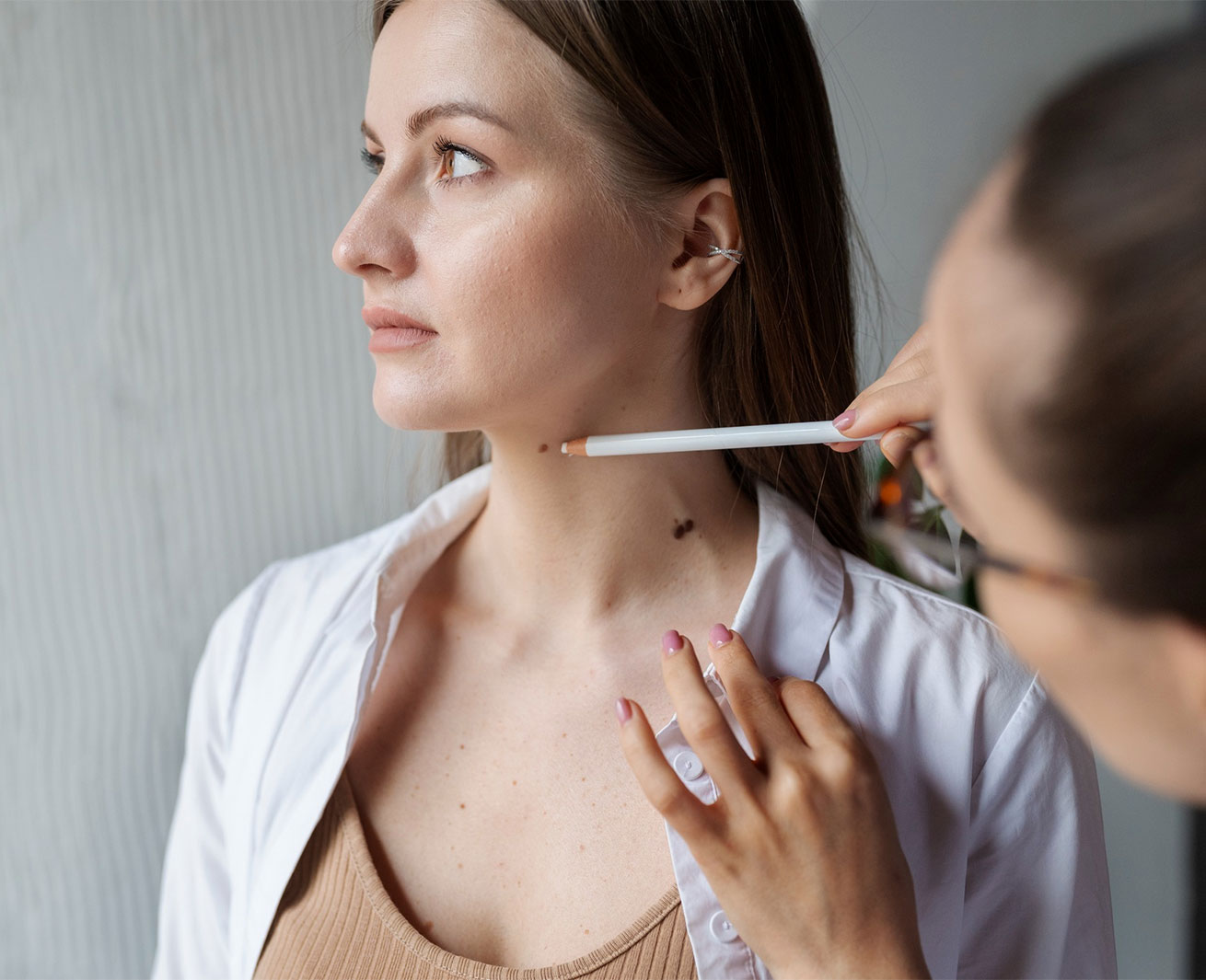
Monitoring of moles is done by dermoscopy (with a special hand lens) and, if necessary, full-body mapping (with digital dermoscopy and full body photography) at least once a year, as well as by training high-risk individuals to self-examine themselves monthly. Indications for biopsy or complete surgical removal of a nevus are the sudden presence of symptoms, such as pain, itching or bleeding, as well as changes in morphological characteristics, such as shape, color, borders, size and symmetry - according to the ABCDE rule (Asymmetry, Border, Color, Diameter, Evolution) -.