- 54 VAS. SOFIAS AVENUE ATHENS 115 28
- (+30) 210 729 75 71
Skin Neoplasm
Melanoma is a form of skin cancer that originates from neoplastic melanocytes. It accounts for only 2% of all skin cancers, but is responsible for the vast majority of deaths, as it tends to metastasize to lymph nodes and internal organs. It affects all ages, with an average age of diagnosis in the 5th and 6th decade of life, while in recent years its incidence has tripled.
The main risk factors for the occurrence of melanoma are overexposure to ultraviolet radiation - especially when it leads to severe sunburns during childhood and adolescence or when it is intense and intermittent -, light skin phototype, genetic predisposition, the presence of a large number of moles on the skin (>100), dysplastic nevus syndrome or, more rarely, a large congenital nevus (>20cm), as well as immunosuppression.
Clinically, a typical melanoma usually appears as a pigmented flat spot with asymmetry, variegation, indistinct contour and a diameter of >0.5cm, which often develops into a plaque on which papules or nodules may present. The most common location is the trunk and extremities.
A lesion suspected of melanoma, which requires further evaluation, can be either a newly appearing nevus with an abnormal appearance and increasing size, or a pre-existing nevus that differs in its characteristics from the rest of the moles on the body or whose appearance has changed noticeably in recent months. Based on the well-known ABCDE rule (Asymmetry, Border, Color, Diameter, Evolution), in a lesion, asymmetry, unclear and irregular borders, uneven distribution of pigment and change in its shape and size are always evaluated.
As a preventive measure, sun protection is essential from childhood, both by avoiding sun exposure as much as possible and by systematically using an appropriate sunscreen as well as an examination in a dermatological clinic at least once a year. In addition, for high-risk individuals, self-examination once a month is recommended, after relevant training by a dermatologist.
A comprehensive approach in the clinic includes taking a detailed personal and family history and clinical evaluation with dermoscopy (using a special hand lens) and full-body mapping of moles (with digital dermoscopy and full-body photography). Early diagnosis and care management of melanoma is always associated with a better prognosis.
The treatment of melanoma is its surgical removal within healthy margins, both circumferentially and in depth, as determined by international protocols. Histological examination of the lesion is particularly important because, on the one hand, it helps in the staging and prognosis of melanoma and, on the other hand, it determines its further therapeutic management. In advanced lesions and in high-risk patients, systemic treatment is recommended (immunotherapy, molecularly targeted therapy or classic chemotherapy and radiotherapy) with excellent results.
Actinic keratosis is a common skin lesion caused by localized dysplasia of the skin cells (partial thickness dysplasia). If neglected, these lesions can lead to full-thickness dysplasia of the skin and develop into squamous cell carcinoma. It is noteworthy that 40-60% of these carcinomas begin as untreated actinic keratosis. In general, the presence of multiple actinic keratosis is considered an indicator of photoaging of the skin and is associated with a high risk of developing skin carcinomas.
Predisposing factors in the development of actinic keratosis are chronic and cumulative exposure to ultraviolet solar radiation, light skin phototype and the presence of immunosuppression.
Usual anatomical locations are the head, face and ears, the neck and sun-exposed parts of the trunk and extremities.
Clinically, at the incipient lesions the skin shows changes in color and texture, and then becomes rougher with accompanying scaling and irritation. A typical actinic keratosis appears as a small superficial papule or plaque, with a red or brown color and scaly surface, often accompanied by itching and roughness.
In the 1st stage of its development, actinic keratosis has less hyperkeratosis, can only be detected by palpation and is characterized as "thin". In the 2nd stage, it is of moderate thickness and is easily visible and palpable, while in the 3rd stage, it has increased hyperkeratosis and is characterized as "thick".
Diagnosis is made through a detailed clinical examination and dermoscopy with a special hand lens, while, in cases where histological confirmation is required, a biopsy is taken.
The proposed treatments are divided into invasive treatments that aim to destroy isolated lesions and medical therapies that have the advantage of being able to treat large areas with many lesions. Invasive treatments include cryotherapy with liquid nitrogen, electrodesiccation and curettage, ablation using CO2 laser devices, and dermabrasion with various special mechanical means (such as wire brushes or diamond wheels). Medical treatments include the topical use of medications (such as 5-fluorouracil, imiquimod and diclofenac), chemical peeling with trichloroacetic acid and photodynamic therapy.
Prevention is primarily based on photoprotection, by avoiding exposure to intense sunlight, proper application of sunscreen and avoiding artificial tanning sources. Secondarily, it is based on early diagnosis of lesions through regular dermatological examination, as well as self-examination for high-risk individuals.
Squamous cell carcinoma is a skin cancer characterized by cellular dysplasia throughout the thickness of the epidermis, but also infiltration of the basement membrane with extension into the underlying dermis. By extending beyond the basement membrane, squamous cell carcinoma acquires metastatic potential and is called invasive. Squamous cell carcinoma is the second most common type of skin cancer after basal cell carcinoma and the second most deadly after melanoma and its incidence in white race appears to be increasing worldwide.
Main predisposing factors for its occurrence are chronic and cumulative exposure to ultraviolet solar radiation, fair skin phototype and immunosuppression.
It is usually found in chronically sun-exposed areas of the body such as the face, head and hands, and often develops on pre-existing actinic keratosis.
Initially, a typical squamous cell carcinoma appears as a red-white hyperkeratotic plaque and after that, as a hard nodule with accompanying ulceration. As the lesion progresses, it causes local tissue destruction and, if not treated promptly, can metastasize.
Diagnosis is based on a detailed clinical examination, dermoscopy with a special hand lens and the histological examination of the lesion by taking a skin biopsy.
The treatment of choice is surgical removal of the lesion, while for low-risk lesions, electrodesiccation and curettage or cryosurgery with liquid nitrogen may be chosen. Additionally, in high-risk or metastatic carcinomas, systemic chemotherapy and immunotherapy, as well as radiotherapy, are used.
Prevention is primarily based on photoprotection by avoiding exposure to intense sunlight, proper application of sunscreen and avoiding artificial tanning sources. Secondarily, it is based on the early detection and treatment of precancerous lesions.
In case there are risk factors for the development of invasive squamous cell carcinoma, dermatological examination is recommended annually for people over 40 years of age and every 3 years for younger people. For patients who have already received treatment for high-risk squamous cell carcinoma, dermatological monitoring is recommended more regularly, as there is a high probability of recurrence or development of a new tumor in another anatomical location.
The prognosis is proportional to the degree of differentiation of the tumor, which is determined histologically. Highly differentiated squamous cell carcinomas have a good prognosis, while poorly differentiated ones have a poor prognosis. It has also been shown that the use of sunscreen reduces the incidence of squamous cell carcinoma and its early treatment can lead to a complete cure.
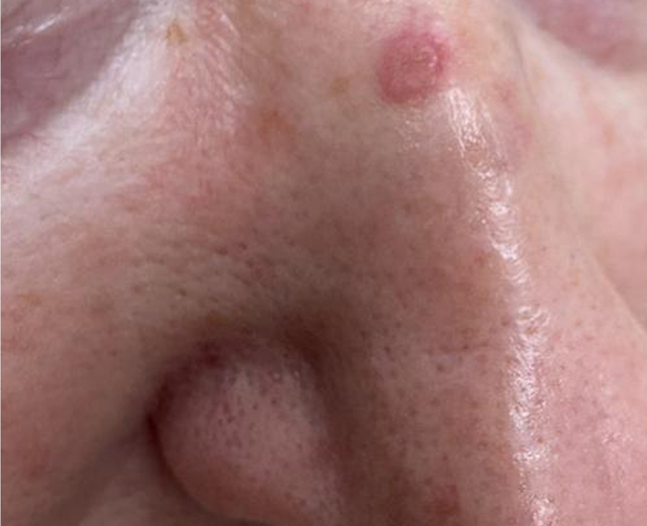
Basal cell carcinoma arises from the basal cells of the epidermis, which are found in the lower layer of the skin. It is a tumor without symptoms that grows slowly and, although it rarely metastasizes, if neglected, it can spread to surrounding tissues destroying them. It is the most common skin cancer and the most common type of cancer in the white race. In the past, it affected middle-aged and elderly people, but in recent years it has also affected younger people.
The main etiological factors for its occurrence are intense sun exposure with a history of severe sunburn during childhood and adolescence, the use of artificial tanning sources, light phototype, immunosuppression and genetic predisposition.
Ιs mainly located on parts of the body exposed to the sun, such as the face, head, neck, back, shoulders and hands.
Clinically, a typical lesion appears as a reddish-pink papule with a shiny appearance and a central erosion or ulceration that bleeds regularly, does not heal and is often covered by a crust. The most common clinical type of basal cell carcinoma is the nodular and ulcerative type, which affects 60% of cases.
Its diagnosis is based on a detailed clinical examination, dermoscopy with a special hand lens and the histological examination of the lesion by taking a skin biopsy.
The treatment of choice is surgical removal of the lesion within healthy margins. Lesions with a low risk of recurrence can be destroyed by local methods such as electrosurgery by curettage and cauterization, cryosurgery using liquid nitrogen and ablation using a CO2 laser device. Other treatment options are photodynamic therapy, immunotherapy, radiotherapy and local or systemic chemotherapy. The choice of treatment is made taking into account the type of lesion, size, location, depth of infiltration, as well as the age and general health status of each patient.
Prevention, primarily, is based on avoiding risk factors, such as solar radiation and artificial tanning, as well as strict sun protection with the correct use of an appropriate sunscreen. Secondarily, it is based on early diagnosis of lesions through monthly self-examination and annual dermatological examination.
Patients who have already received treatment for basal cell carcinoma are considered to be at high risk of developing another type of skin cancer or developing basal cell carcinoma in another anatomical location. For this reason, routine dermatological examination is recommended to be more regular.
The prognosis of basal cell carcinoma is excellent if treated promptly, but, if neglected, its local spread can cause significant morbidity and cosmetic disfigurement.